Δευ: 18:30 – 21:30 - Τετ:18:30 – 21:30 - Παρ:18:30 – 21:30
Δερβενακίων 30 & Υγείας, Παλλήνη
It is the result of prolonged injury during inversion of the ankle. It is characterized by necrosis of a portion of bone and articular cartilage, which can be separated from the ankle vault. The necrosis can develop on the medial side of the ankle, due to compression of the medial side of the ankle vault by the tibia or on the lateral side, as a result of forces exerted by the fibula. The internal lesions are posterior, while the external, more anterior Osteochondritis of the ankle Osteochondritis of the ankle
The symptoms can be acute or chronic. The child usually has diffuse pain in the ankle and swelling. If an acute injury has preceded, its symptoms predominate, subside after 3-6 weeks and are followed by chronic symptoms. Otherwise, there are chronic symptoms (pain, stiffness, instability or blockage) with/or without a history of acute injury.
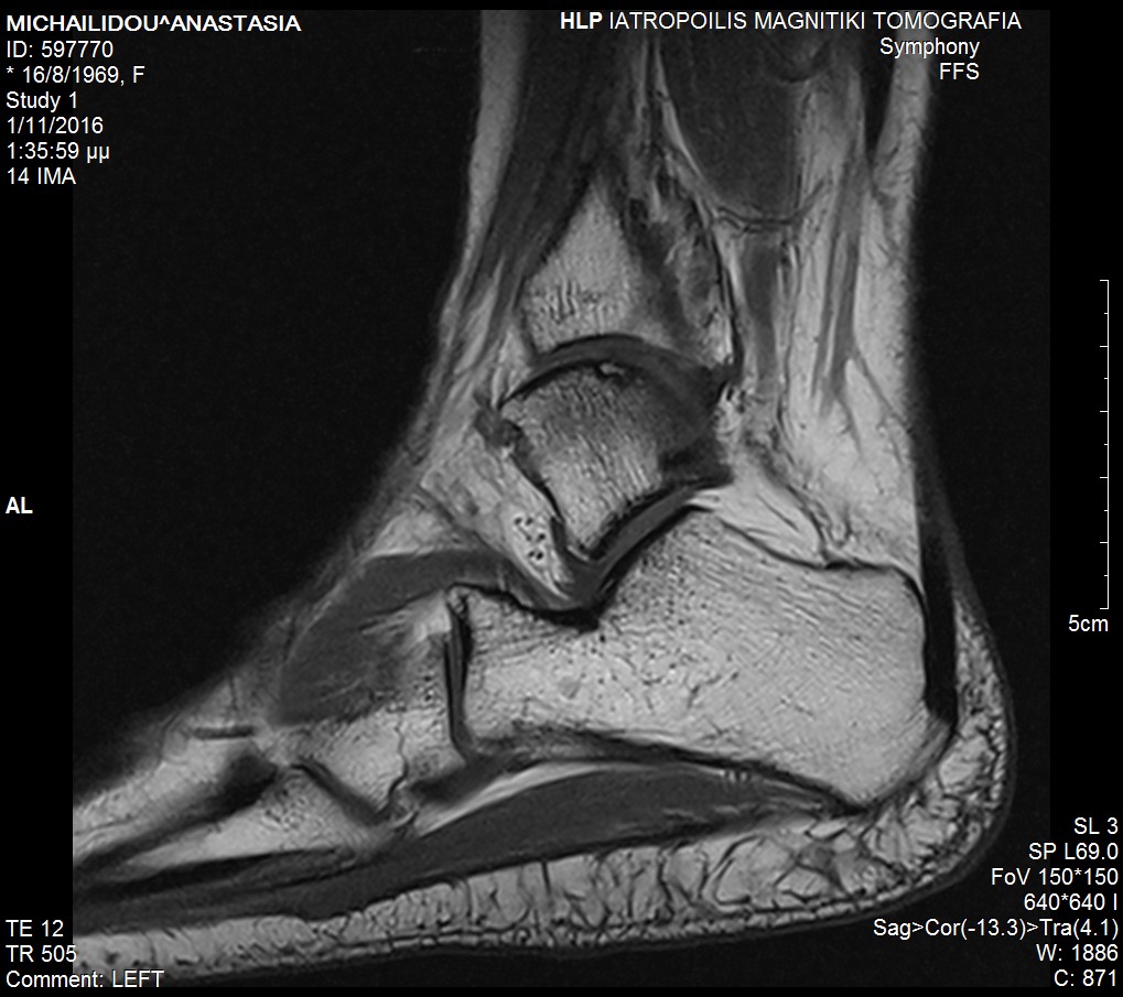
In the early stages, diagnosis is difficult and the disease is usually treated as a sprain. It is confirmed with tomography with/or without arthrography or CT scan.
Without effective treatment, this osteochondral damage will result in a painful joint as a result of the development of degenerative osteoarthritis.
The evaluation and staging of the disease is done with the help of special X-rays and Magnetic Resonance Imaging (MRI arthrogram).
Conservative treatment includes immobilization of the joint for at least 8-10 weeks, walking with partial weight-bearing and re-evaluation.
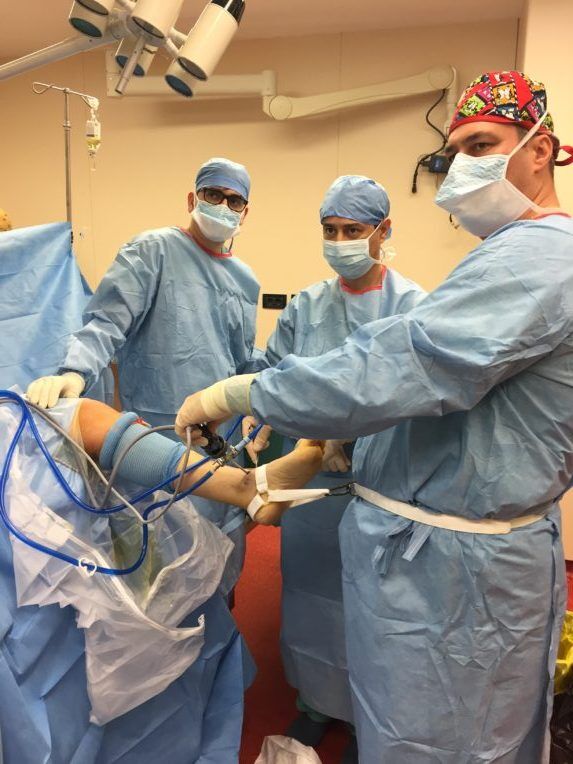
Surgical treatment consists of arthroscopic refixation of the partially or completely detached part using very thin absorbable pins, chondroplasty using the microfracture technique, while when this is not possible, the use of a graft is required to cover the defect (the technique usually requires osteotomy of the medial malleolus and mosaicplasty).
[instagram-feed feed=1]
 copy")
